
Scientists study the differences in brain chemistry between those with and without HD
In a new neuroimaging study, investigators led by Taro Mizobe, Department of Neuropsychiatry, Graduate School of Medical Sciences, Kyushu University, Fukuoka, Japan, compared the brain scans of individuals with and without hoarding disorder (HD) to see if imaging would offer some insight into why some individuals engage in this disorder. Their findings were published online January 18 in the Journal of Psychiatric Research.
Individuals who suffer from hoarding disorder have difficulty getting rid of or possessions due to a perceived need to save the items. These items can be of little or no use or value and yet the individual will still find value in them. Individuals have also been known to hoard plants and animals. Attempts to part with these possessions creates significant distress. Psychological intervention is commonly needed, and recovery is lifelong.
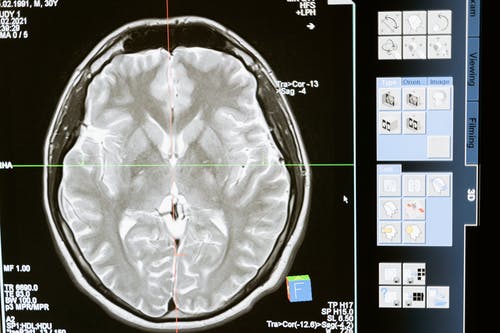
For the current study, participants underwent MRI and DTI scans. The images of 25 individuals with hoarding disorder (mean age, 43 years; 64% women; 96% right-handed) were compared with those of 36 healthy controls. Participants with HD had higher scores on the Hamilton Rating Scales for depression and anxiety than those without HD (P < .001 for both), according to the team.
The results of their study showed that compared with healthy individuals, participants with HD had anatomically widespread abnormalities in WM tracts. Specifically, “a wide range of abnormalities were found in frontal WM related to HD symptom severity, as well as cortical regions involved in cognitive dysfunction,” the team reported.
“The finding of a characteristic association between alterations in the prefrontal WM tract, which connects cortical regions involved in cognitive function and the severity of hoarding symptoms, could provide new insights into the neurobiological basis of HD,” the researchers wrote, adding, “Although there are no clear neurobiological models of HD, several neuroimaging studies have found specific differences in specific brain regions between patients with and without HD.”
Structural MRI studies and voxel-based morphometry have shown larger volumes of gray matter in several regions of the brain in patients with HD. And “diffusion tensor imaging (DTI) studies have yielded inconsistent” findings, therefore little is known about the microstructure of WM in the brains of patients with HD,” they add.
The current study was designed “to investigate microstructural alterations in the WM tracts of individuals with HD” by using tract-based spatial statistics, which is a model typically used for “whole-brain, voxel-wise analysis of DTI measures…DTI neuroimaging can assess the microstructure of WM. In the current study, the investigators focused on the three measures yielded by DTI: fractional anisotropy (FA), which is an index of overall WM integrity; axial diffusivity (AD); and radial diffusivity (RD).”
Of the patients with HD, 10 were taking psychiatric medications such as antidepressants, tranquilizers, or nonstimulant agents for attention deficit hyperactivity disorder (ADHD).
The American Psychiatric Association reports that the overall prevalence of hoarding disorder is approximately 2.6%, with higher rates for people over 60 years old and people with other mental health diagnoses, including anxiety and depression. Evidence shows that trauma can set off HD and that there are relatively equal rates of the disorder in both men and women.
Join the conversation!